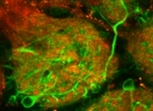
 Using mouse olfactory bulb as a model system we compare the performance of genetically-encoded calcium sensor TN-XXL and small molecule calcium indicators; describe how to choose the right calcium indicator and how to load it into the cells of interest; discuss the use of cell type-specific markers and, finally, illustrate the application of this technique for high resolution in vivo imaging of sensory-driven neuronal activity.
For more details see:
In vivo functional imaging of the olfactory bulb at single cell resolution, S, Fink, Y Kovalchuk, R Homma, B Schwendele, S Direnberger, LB Cohen, O Griesbeck, and O Garaschuk, Neuronal Network Analysis (in Press)