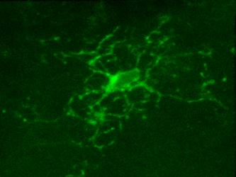
 Microglia, resident immune cells of the brain, react to the presence of pathogens/danger signals with a large repertoire of functional responses including morphological changes, proliferation, chemotaxis, production/release of cytokines, and phagocytosis. In vitro studies suggest that many of these effector functions are Ca2+-dependent, but our knowledge about in vivo Ca2+ signalling in microglia is rudimentary. This is mostly due to technical reasons, as microglia largely resisted all attempts of in vivo labelling with Ca2+ indicators. Here, we introduce a novel approach, utilizing a microglia-specific microRNA-9-regulated viral vector, enabling the expression of a genetically-encoded ratiometric Ca2+ sensor Twitch-2B in microglia.
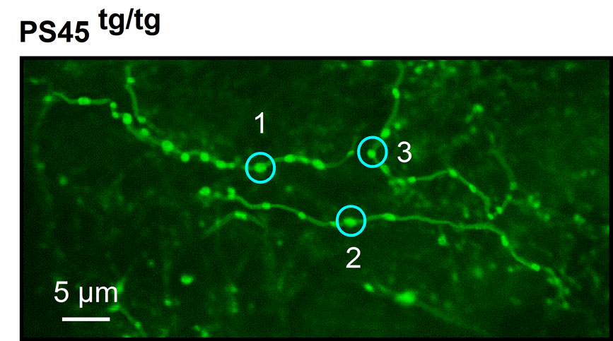 Neuronal hyperactivity is the emerging functional hallmark of Alzheimer’s disease (AD) in both humans and different mouse models, mediating an impairment of memory and cognition. The mechanisms underlying neuronal hyperactivity remain, however, elusive. In vivo Ca2+ imaging of somatic, dendritic, and axonal activity patterns of cortical neurons revealed that both healthy aging and AD-related mutations augment neuronal hyperactivity. The AD-related enhancement occurred even without amyloid deposition and neuroinflammation, mainly due to presenilin-mediated dysfunction of intracellular Ca2+ stores in presynaptic boutons, likely causing more frequent activation of synaptic NMDA receptors.
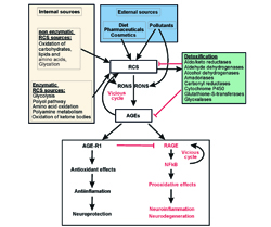 Brains’ high energy expenditure with preferable utilization of glucose and ketone bodies, defines the specific features of its energy homeostasis. The extensive oxidative metabolism is accompanied by a concomitant generation of high amounts of reactive oxygen, nitrogen, and carbonyl species, which will be here collectively referred to as RONCS. Such metabolism in combination with high content of polyunsaturated fatty acids creates specific problems in maintaining brains’ redox homeostasis. While the levels of products of interaction between RONCS and cellular components increase slowly during the first two trimesters of individuals’ life, their increase is substantially accelerated towards the end of life.
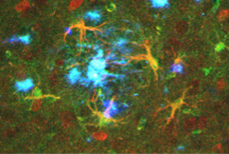 Functioning at the interface between the nervous and immune systems, in the amyloid-depositing brain, astrocytes become hypertrophic and accumulate around senile plaques. Moreover, hippocampal astrocytes upregulate their γ-aminobutyric acid (GABA) content and enhance tonic inhibition, likely causing local circuit imbalance. It remains, however, unclear whether this effect is hippocampus specific and how it is regulated during disease progression. Here, we studied changes in astrocytic morphology and GABA content in the frontal cortex and dentate gyrus of control and amyloid-depositing mice. Healthy aging was accompanied by a transient increase in astrocytic GABA content at middle age and region-specific alterations of soma size.
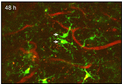 Microglia play key roles in brain development, homeostasis, and function, and it is widely assumed that the adult population is long lived and maintained by self-renewal. However, the precise temporal and spatial dynamics of the microglial population are unknown. We show in mice and humans that the turnover of microglia is remarkably fast, allowing the whole population to be renewed several times during a lifetime. The number of microglial cells remains steady from late postnatal stages until aging and is maintained by the spatial and temporal coupling of proliferation and apoptosis, as shown by pulse-chase studies, chronic in vivo imaging of microglia, and the use of mouse models of dysregulated apoptosis.
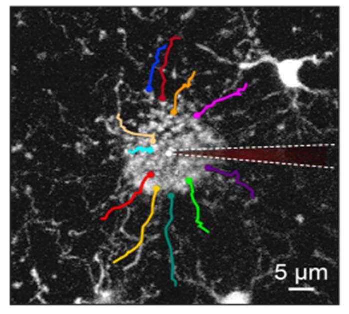
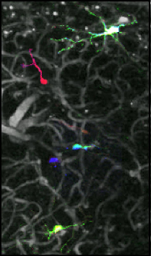 The behavior of adult-born cells can be easily monitored in cell culture or in lower model organisms, but longitudinal observation of individual mammalian adult-born cells in their native microenvironment still proves to be a challenge. Here we have established an approach named optical cell positioning system for long-term in vivo single-cell tracking, which integrates red-green-blue cell labeling with repeated angiography. By combining this approach with in vivo two-photon imaging technique, we characterized the in vivo migration patterns of adult-born neurons in the olfactory bulb.
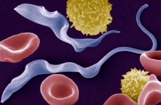
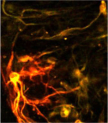 Astrocytic brain tumours, including glioblastomas, are incurable neoplasms characterized by diffusely infiltrative growth. Here we show that many tumour cells in astrocytomas extend ultra-long membrane protrusions, and use these distinct tumour microtubes as routes for brain invasion, proliferation, and to interconnect over long distances. The resulting network allows multicellular communication through microtube-associated gap junctions. When damage to the network occurred, tumour microtubes were used for repair. Moreover, the microtube-connected astrocytoma cells, but not those remaining unconnected throughout tumour progression, were protected from cell death inflicted by radiotherapy.
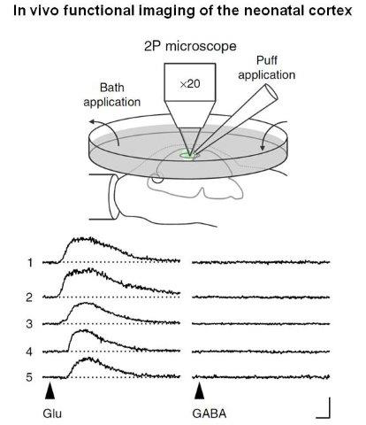 A large body of evidence from in vitro studies suggests that GABA is depolarizing during early postnatal development. However, the mode of GABA action in the intact developing brain is unknown. Here we examine the in vivo effects of GABA in cells of the upper cortical plate using a combination of electrophysiological and Ca2+-imaging techniques. We report that at postnatal days (P) 3-4, GABA depolarizes the majority of immature neurons in the occipital cortex of anaesthetized mice. At the same time, GABA does not efficiently activate voltage-gated Ca2+ channels and fails to induce action potential firing. Blocking GABAA receptors disinhibits spontaneous network activity, whereas allosteric activation of GABAA receptors has the opposite effect.