 Astrocytic brain tumours, including glioblastomas, are incurable neoplasms characterized by diffusely infiltrative growth. Here we show that many tumour cells in astrocytomas extend ultra-long membrane protrusions, and use these distinct tumour microtubes as routes for brain invasion, proliferation, and to interconnect over long distances. The resulting network allows multicellular communication through microtube-associated gap junctions. When damage to the network occurred, tumour microtubes were used for repair. Moreover, the microtube-connected astrocytoma cells, but not those remaining unconnected throughout tumour progression, were protected from cell death inflicted by radiotherapy.
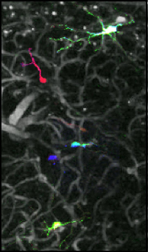 The behavior of adult-born cells can be easily monitored in cell culture or in lower model organisms, but longitudinal observation of individual mammalian adult-born cells in their native microenvironment still proves to be a challenge. Here we have established an approach named optical cell positioning system for long-term in vivo single-cell tracking, which integrates red-green-blue cell labeling with repeated angiography. By combining this approach with in vivo two-photon imaging technique, we characterized the in vivo migration patterns of adult-born neurons in the olfactory bulb.
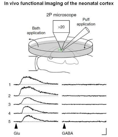 A large body of evidence from in vitro studies suggests that GABA is depolarizing during early postnatal development. However, the mode of GABA action in the intact developing brain is unknown. Here we examine the in vivo effects of GABA in cells of the upper cortical plate using a combination of electrophysiological and Ca2+-imaging techniques. We report that at postnatal days (P) 3-4, GABA depolarizes the majority of immature neurons in the occipital cortex of anaesthetized mice. At the same time, GABA does not efficiently activate voltage-gated Ca2+ channels and fails to induce action potential firing. Blocking GABAA receptors disinhibits spontaneous network activity, whereas allosteric activation of GABAA receptors has the opposite effect.
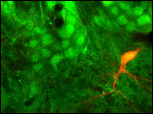 Juxtaglomerular neurons (JGNs) of the mammalian olfactory bulb are generated throughout life. Their integration into the preexisting neural network, their differentiation and survival therein depend on sensory activity, but when and how these adult-born cells acquire responsiveness to sensory stimuli remains unknown. In vivo two-photon imaging of retrovirally labelled adult-born JGNs reveals that ~90% of the cells arrive at the glomerular layer after day post injection (DPI) 7. After arrival, adult-born JGNs are still migrating, but at DPI 9, 52% of them have odour-evoked Ca(2+) signals. Their odourant sensitivity closely resembles that of the parent glomerulus and surrounding JGNs, and their spontaneous and odour-evoked spiking is similar to that of their resident neighbours.
 The dynamics of β-amyloid deposition and related second-order physiological effects, such as regional cerebral blood flow (rCBF), are key factors for a deeper understanding of Alzheimer's disease (AD). We present longitudinal in vivo data on the dynamics of β-amyloid deposition and the decline of rCBF in two different amyloid precursor protein (APP) transgenic mouse models of AD. Using a multiparametric positron emission tomography and magnetic resonance imaging approach, we demonstrate that in the presence of cerebral β-amyloid angiopathy (CAA), β-amyloid deposition is accompanied by a decline of rCBF. Loss of perfusion correlates with the growth of β-amyloid plaque burden but is not related to the number of CAA-induced microhemorrhages.
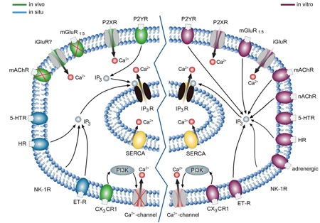
 Neuroinflammation is a hallmark of Alzheimer’s disease (AD) both in man and in multiple mouse models, and epidemiological studies link the use of anti-inflammatory drugs with a reduced risk of developing the disease. AD-related neuroinflammation is largely mediated by microglia, the main immune cells of the central nervous system. In vitro, executive functions of microglia are regulated by intracellular Ca2+ signals, but little is known about microglial Ca2+ signaling in vivo. Here we analyze in vivo properties of these cells in two mouse models of AD. In both strains plaque-associated microglia had hypertrophic/amoeboid morphology and were strongly positive for markers of activation such as CD11b and CD68.
 The quality of genetically encoded calcium indicators (GECIs) has improved dramatically in recent years, but high-performing ratiometric indicators are still rare. Here we describe a series of fluorescence resonance energy transfer (FRET)-based calcium biosensors with a reduced number of calcium binding sites per sensor. These 'Twitch' sensors are based on the C-terminal domain of Opsanus troponin C. Their FRET responses were optimized by a large-scale functional screen in bacterial colonies, refined by a secondary screen in rat hippocampal neuron cultures. We tested the in vivo performance of the most sensitive variants in the brain and lymph nodes of mice.
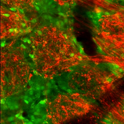 Juxtaglomerular neurons represent one of the largest cellular populations in the mammalian olfactory bulb yet their role for signal processing remains unclear. We used two-photon imaging and electrophysiological recordings to clarify the in vivo properties of these cells and their functional organization in the juxtaglomerular space. Juxtaglomerular neurons coded for many perceptual characteristics of the olfactory stimulus such as (1) identity of the odorant, (2) odorant concentration, (3) odorant onset, and (4) offset.