Live imaging of microglia during sleeping sickness reveals early and heterogeneous inflammatory responses
Invasion of the central nervous system (CNS) is the most serious consequence of Trypanosoma brucei infection, which causes sleeping sickness. Recent experimental data have revealed some more insights into the disease during the meningoencephalitic stage. However, detailed cellular processes befalling the CNS during the disease are poorly understood. To further address this issue, we implanted a cranial window on the cortex of B6.129P2(Cg)-Cx3cr1tm1Litt/J mice, infected them with Trypanosoma brucei expressing RFP via intraperitoneal injection, and monitored microglial cells and parasites longitudinally over 30 days using in vivo 2-photon imaging. We correlated the observed changes with histological analyses to evaluate the recruitment of peripheral immune cells. We uncovered an early involvement of microglia that precedes invasion of the CNS by the parasite.
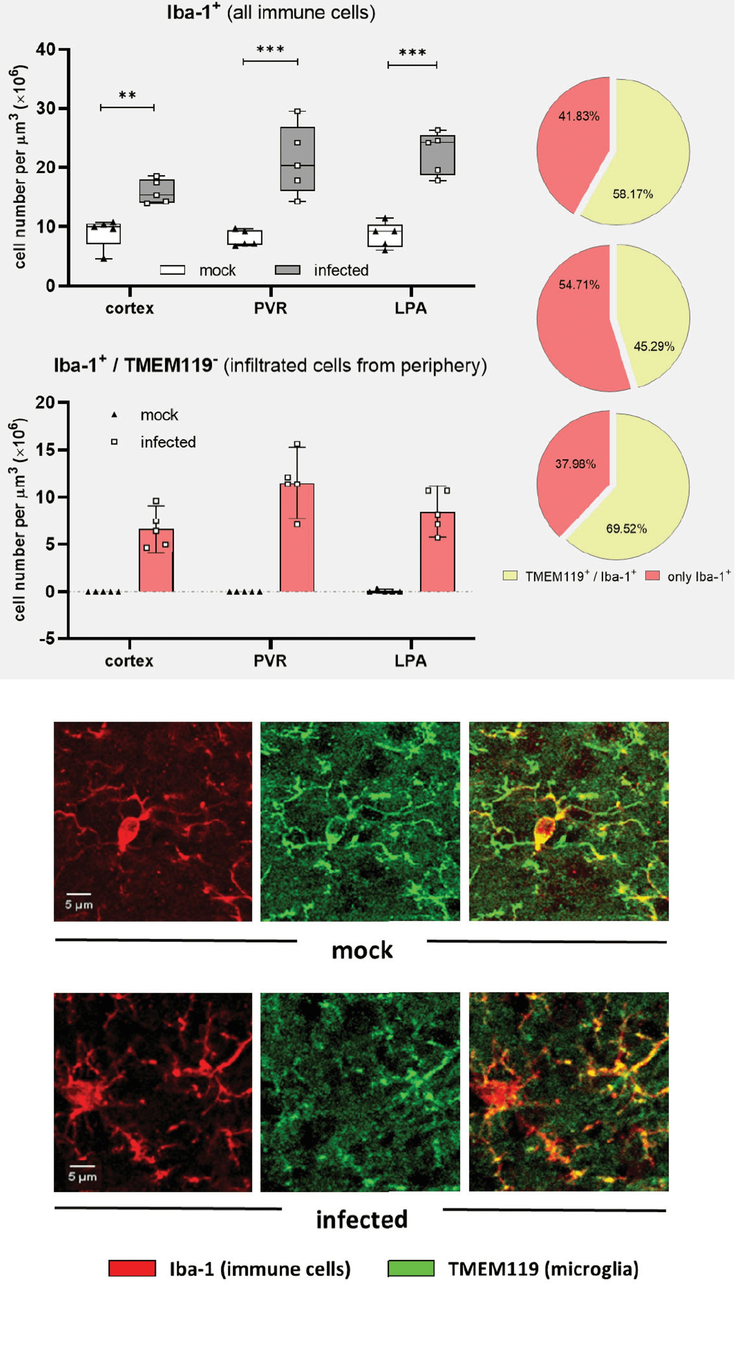
We accomplished a detailed characterization of the progressive sequence of events that correlates with microglial morphological changes and microgliosis. Our findings unveiled a heterogeneous microglial response in places of initial homeostatic disruption near brain barriers and pointed out an exceptional capability of microglia to hamper parasite proliferation inside the brain. We also found early signs of inflammation in the meninges, which synchronize with the microglial response. Moreover, we observed a massive infiltration of peripheral immune cells into the parenchyma as a signature in the final disease stage. Overall, our study provides new insights into the host-pathogen immune interactions in the meningeal and parenchymal compartments of the neocortex.
The article is accessible on the following DOI: https://doi.org/10.3389/fimmu.2023.1253648