 Microglia, the resident immune cells of the central nervous system, exhibit a wide array of functional states, even in their so-called “homeostatic” condition, when they are not actively responding to overt pathological stimuli. These functional states can be visualized using a combination of multi-omics techniques (e.g., gene and protein expression, posttranslational modifications, mRNA profiling, and metabolomics), and, in the case of homeostatic microglia, are largely defined by the global (e.g., genetic variations, organism’s age, sex, circadian rhythms, and gut microbiota) as well as local (specific area of the brain, immediate microglial surrounding, neuron-glia interactions and synaptic density/activity) signals (Paolicelli et al., 2022).
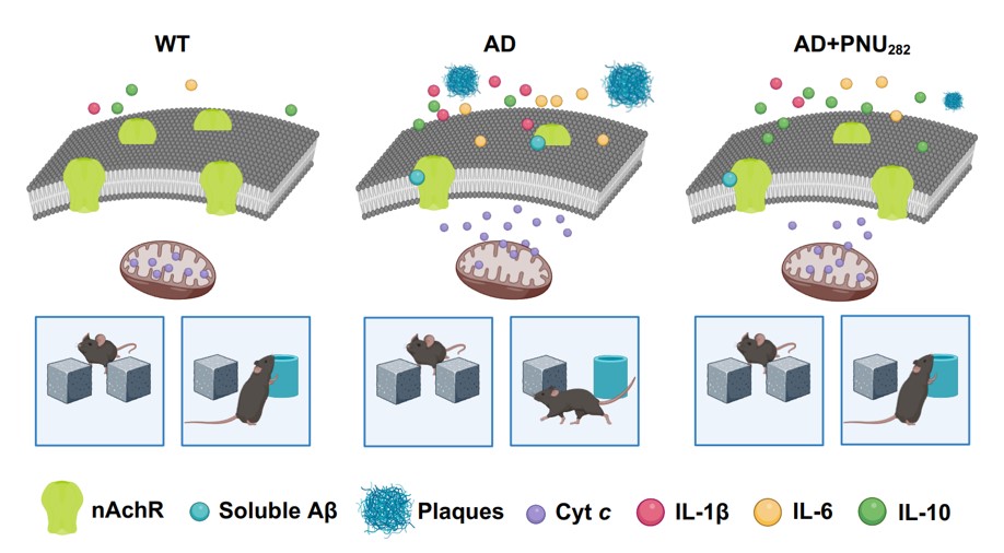 Alzheimer's disease (AD) is an age-dependent incurable neurodegenerative disorder accompanied by neuroinflammation, amyloid accumulation, and memory impairment. It begins decades before the first clinical symptoms appear, and identifying early biomarkers is key for developing disease-modifying therapies. We show now in a mouse model of AD that before any amyloid deposition the brains of 1.5-month-old mice contain increased levels of pro-inflammatory cytokines IL-1β and IL-6, decreased levels of nicotinic acetylcholine receptors (nAChRs) in the brain and brain mitochondria and increased amounts of α7 nAChR-bound Aβ1-42, along with impaired episodic memory and increased risk of apoptosis.
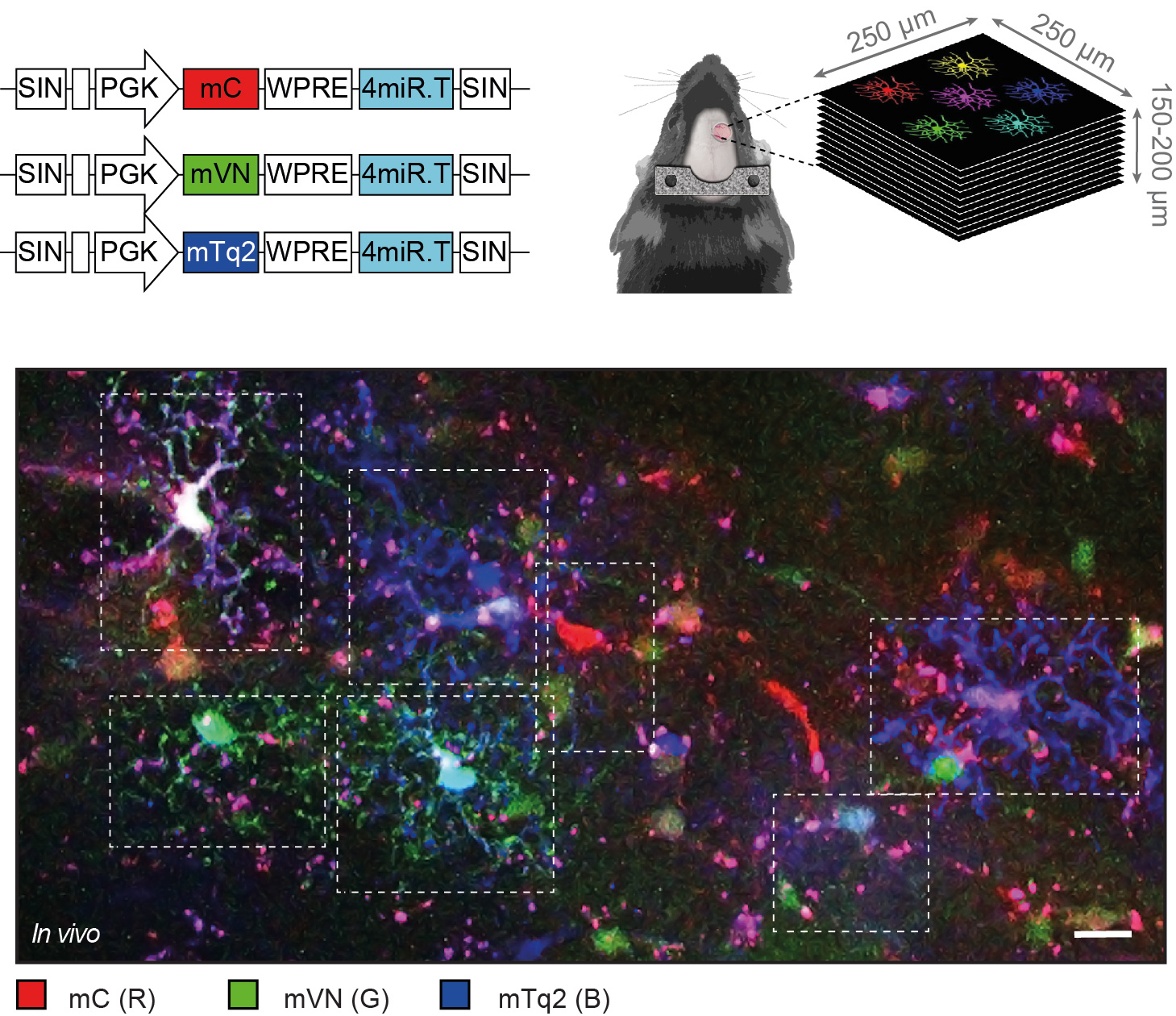
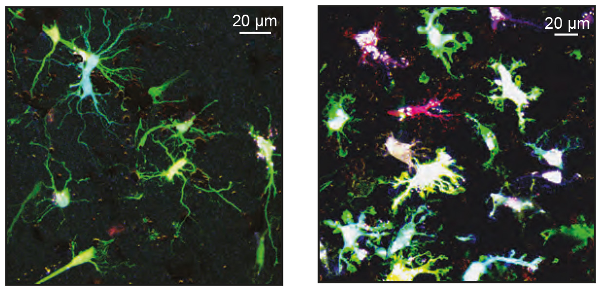 Key functions of Ca2+ signaling in rodent microglia include monitoring the brain state as well as the surrounding neuronal activity and sensing the danger or damage in their vicinity. Microglial Ca2+ dyshomeostasis is a disease hallmark in many mouse models of neurological disorders but the Ca2+ signal properties of human microglia remain unknown. We developed a novel genetically-encoded ratiometric Ca2+ indicator, targeting microglial cells in the freshly resected human tissue, organotypically cultured tissue slices and analyzed in situ ongoing Ca2+ signaling of decades-old microglia dwelling in their native microenvironment.
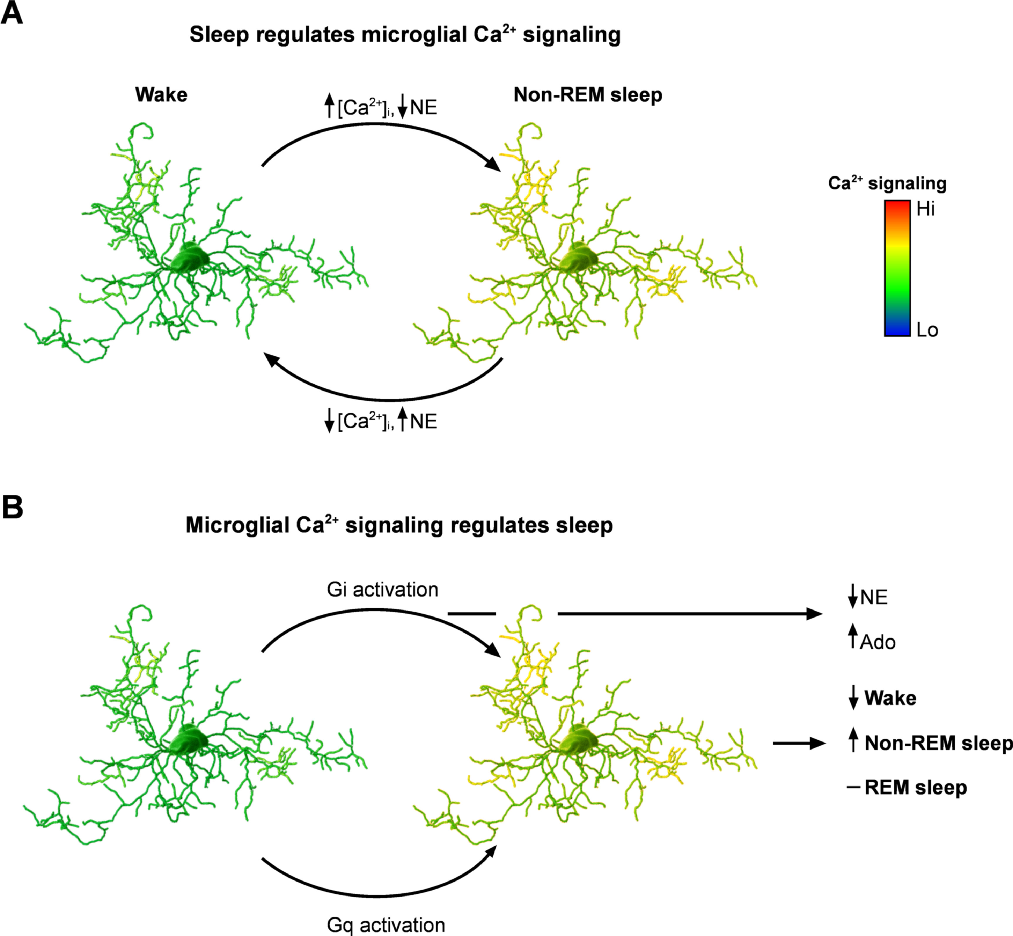 Why evolution made sleep an almost ubiquitous property of animals remains an enigma; similarly, the mechanisms regulating the sleep-wake cycle, although being extensively studied over decades remain controversial. The central role of neuroglia in sleep was proposed by Santiago Ramón y Cajal in 1895. He postulated that fine processes of astrocytes can insert in-between synaptic contacts thus limiting information transfer and instigating sleep.
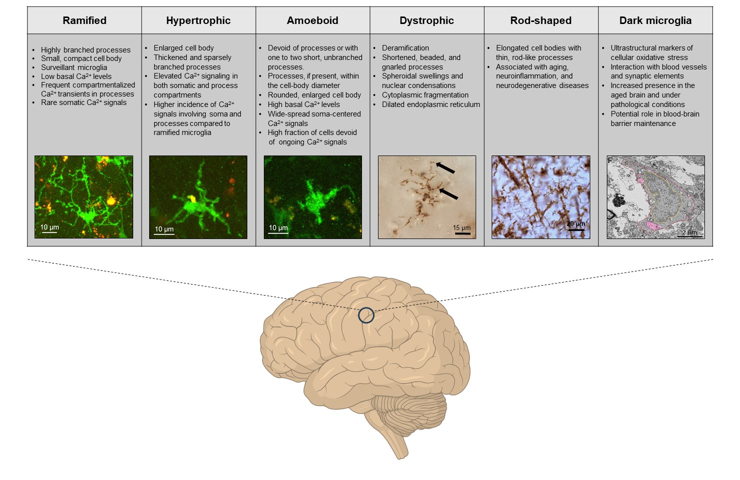
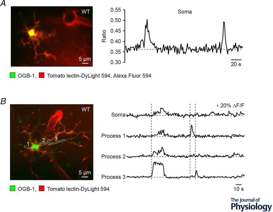 Under physiological conditions microglia, the immune sentinels of the brain, constantly monitor their microenvironment. In the case of danger, damage or cell/tissue dyshomeostasis, they react with changes in process motility, polarization, directed process movement, morphology and gene expression profile; release pro- and anti-inflammatory mediators; proliferate; and clean brain parenchyma by means of phagocytosis.
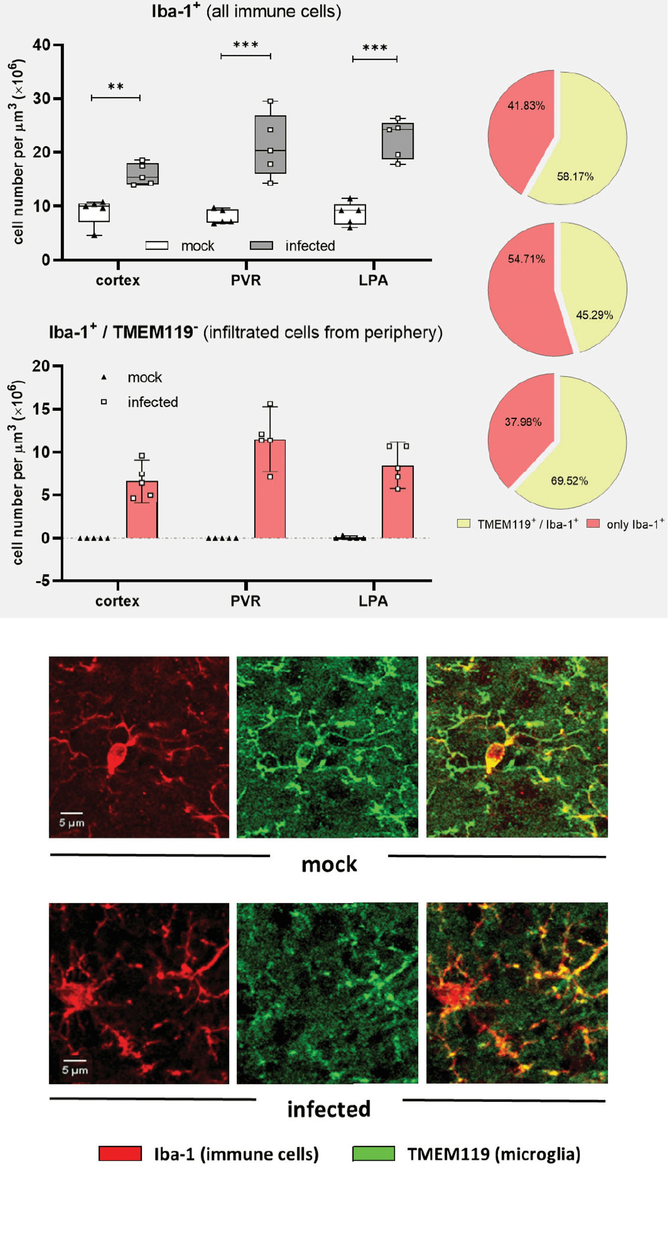
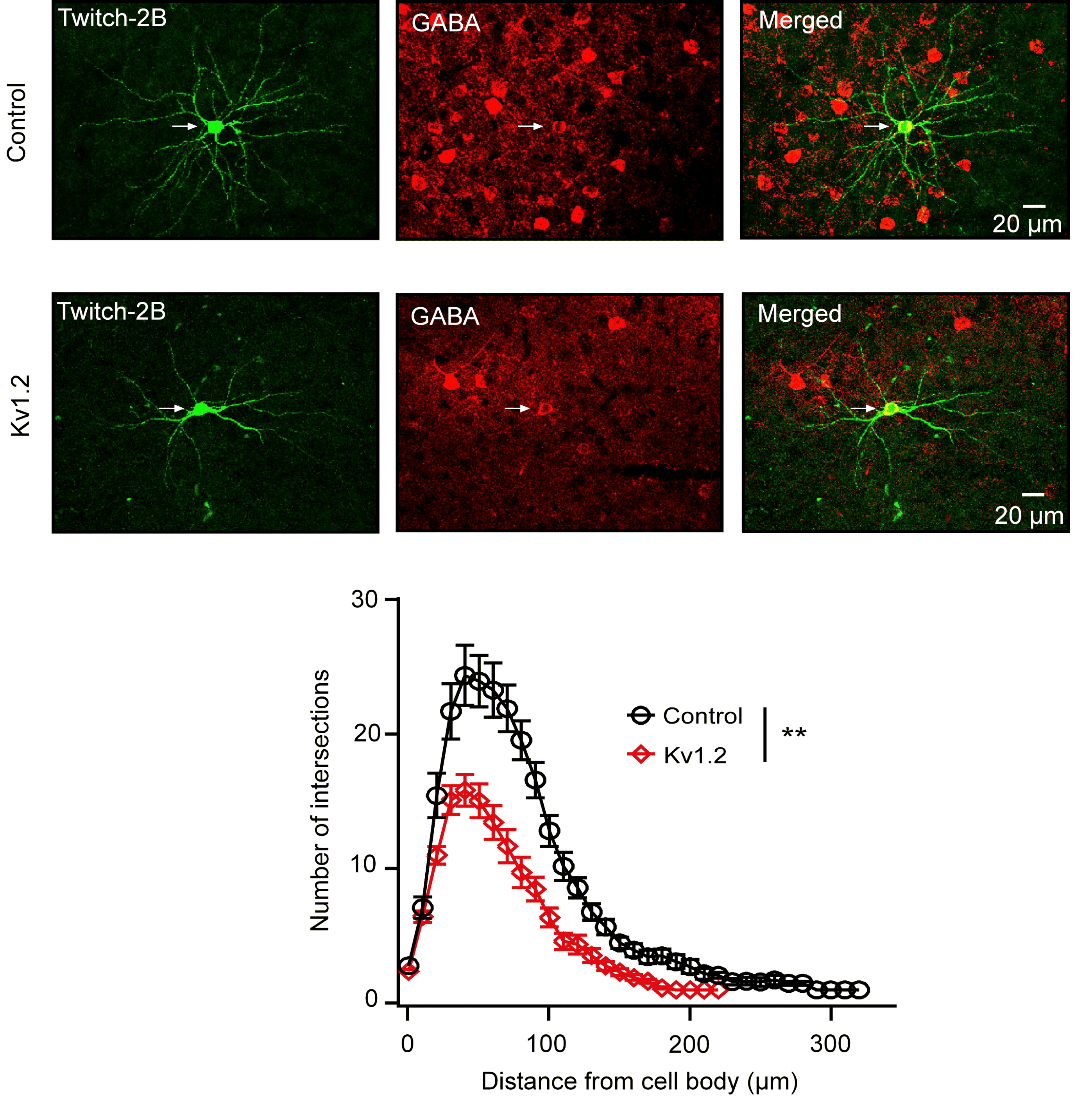 De novo mutations in genes encoding K+ channels are implicated in many severe neurodevelopmental disorders. Specifically, mutations in KCNA2, encoding the Shaker-type voltage-gated K+ channel Kv1.2, and KCNJ2, encoding the inwardly rectifying K+ channel Kir2.1, associate with focal and generalized epilepsies, brain atrophy, autism, ataxia and hereditary spastic paraplegia (Syrbe et al., 2015; Masnada et al., 2017; Cheng et al., 2021).