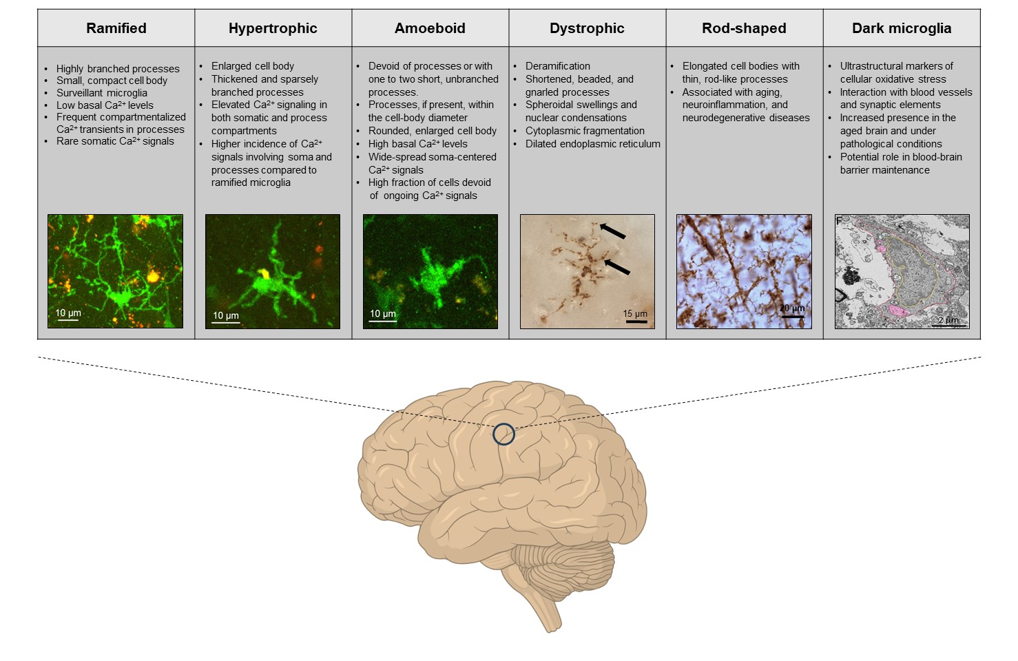
 Microglia, the resident immune cells of the central nervous system, exhibit a wide array of functional states, even in their so-called “homeostatic” condition, when they are not actively responding to overt pathological stimuli. These functional states can be visualized using a combination of multi-omics techniques (e.g., gene and protein expression, posttranslational modifications, mRNA profiling, and metabolomics), and, in the case of homeostatic microglia, are largely defined by the global (e.g., genetic variations, organism’s age, sex, circadian rhythms, and gut microbiota) as well as local (specific area of the brain, immediate microglial surrounding, neuron-glia interactions and synaptic density/activity) signals (Paolicelli et al., 2022).

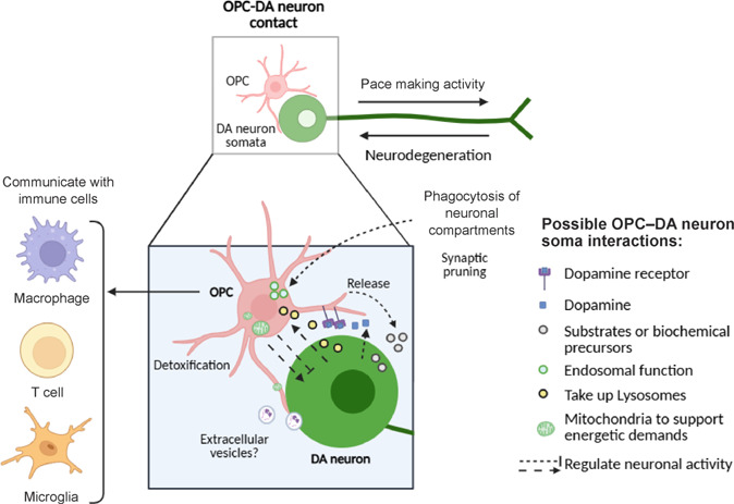 Oligodendroglial cells comprise a large cell population in the substantia nigra (SN). We recently reported a stable portion of oligodendrocyte precursor cells (OPCs) in the SN pars compacta (SNpc) in a 1:1 ratio with dopaminergic neurons: 15% of cells in neonate and young mice, rising to 20% in aged mice. Moreover, this portion represents only 45% of all oligodendroglial cells in the SNpc and 54% of all oligodendroglial cells in the SN pars reticulata (SNpr) (Fitzgerald et al., 2025). The SN is a deeply located area of the midbrain and the site of dopaminergic degeneration in Parkinson’s disease (PD).
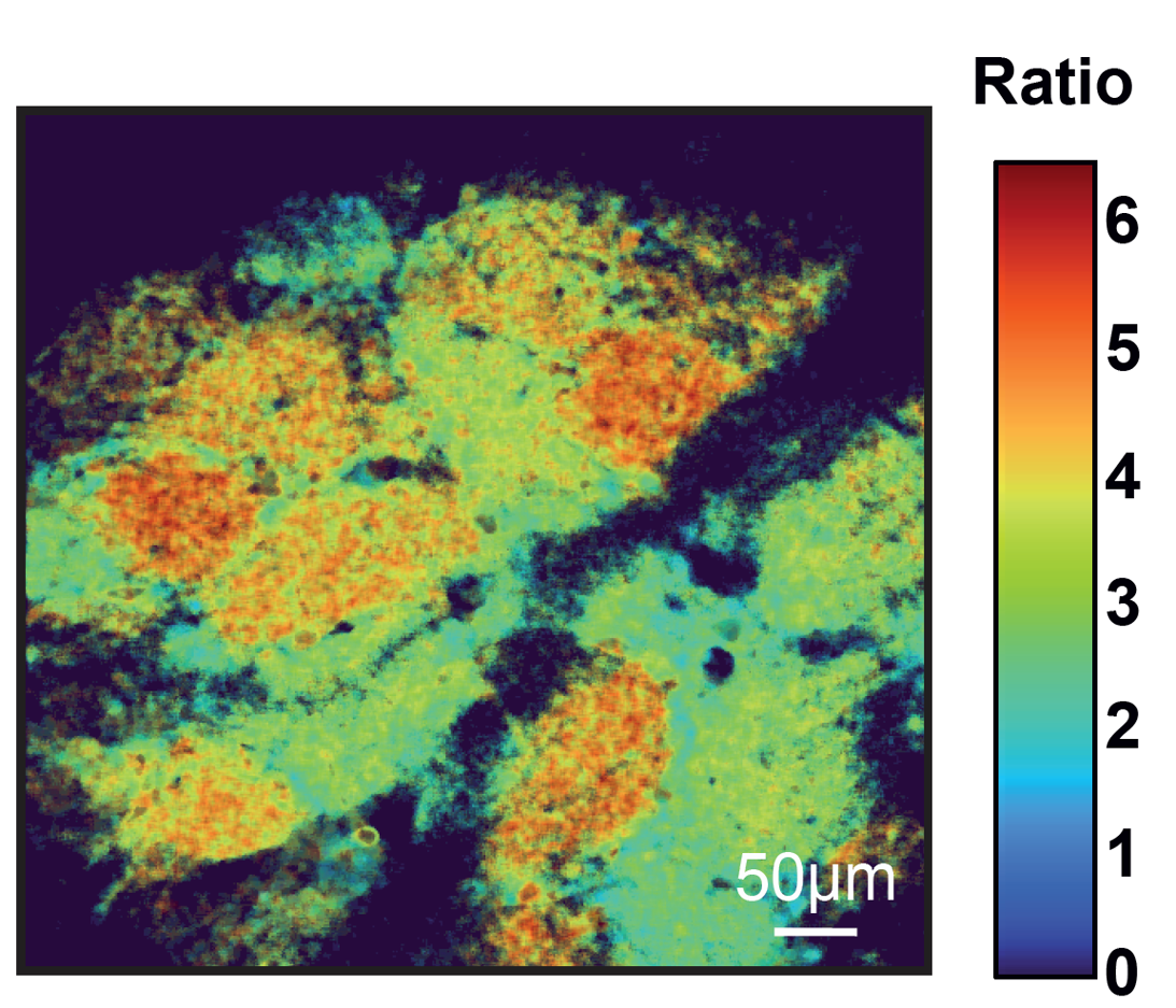 Glomeruli are signal-processing units of the olfactory bulb (OB) that play a key role in many OB computations, including contrast enhancement, gain control, and odorant-selective habituation. In awake mice, we unveil an extremely stable, inhomogeneous map of basal glomerulus-specific activity that serves as the background against which olfactory signal processing occurs. This activity is strongly driven by centrifugal cholinergic inputs; endogenous and airflow-evoked spiking of olfactory sensory neurons; and, to a minor extent, by the odor environment. Moreover, it is brain-state dependent and suppressed under various forms of anesthesia, and is therefore likely attenuated during sleep.
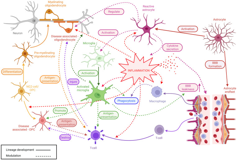 Neuroglial cells are pivotal regulators of innate and adaptive immune processes within the brain and spinal cord. Neuroglia, comprising microglia, astrocytes, oligodendrocytes, oligodendrocyte precursor cells (OPCs), and ependymal cells orchestrate a complex network of immune surveillance, signaling, and blood–brain barrier maintenance, thereby controlling immune cell entry. Microglia function as the resident macrophage-like cells of the central nervous system (CNS) with numerous physiological functions. Microglia continuously monitor the neural environment and rapidly respond to injury, infection, and perturbations in homeostasis, with changes in process motility, polarization, directed process movement, morphology, and gene expression profiles.
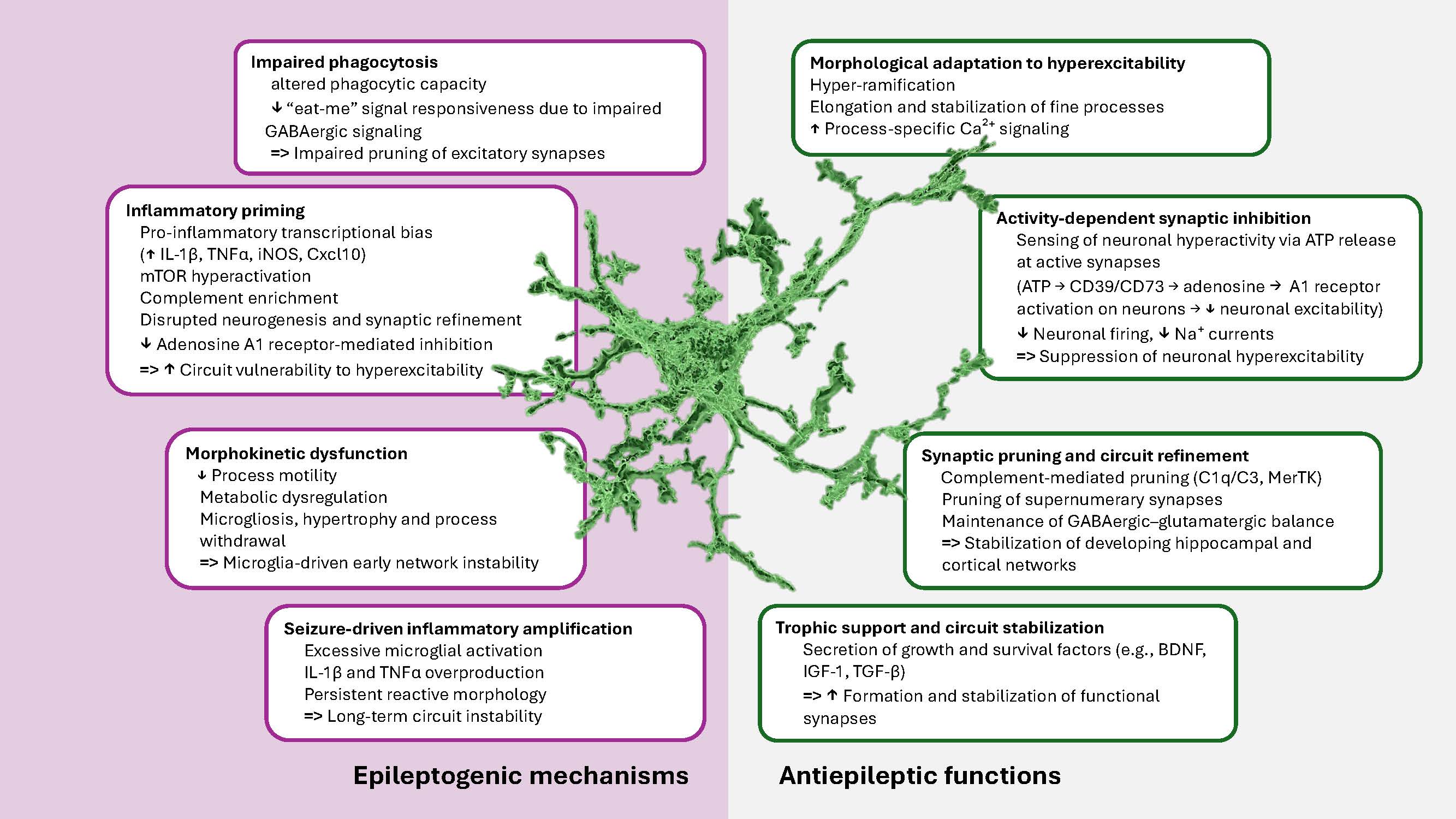 As the brain’s resident macrophages, microglia on the one side are increasingly recognized as essential players in discrete developmental stages, where immune, metabolic, and activity-derived signals are coordinately integrated to guide brain development. On the other side, the precise temporal and molecular coordination of microglial maturation is imperative for the structural and functional integrity of the developing central nervous system (CNS). In this review, we synthesize recent data that reposition microglia from a uniform population of immune sentinels to temporally programmed and regionally specialized regulators of circuit maturation.
 Alzheimer's disease (AD) is an age-dependent incurable neurodegenerative disorder accompanied by neuroinflammation, amyloid accumulation, and memory impairment. It begins decades before the first clinical symptoms appear, and identifying early biomarkers is key for developing disease-modifying therapies. We show now in a mouse model of AD that before any amyloid deposition the brains of 1.5-month-old mice contain increased levels of pro-inflammatory cytokines IL-1β and IL-6, decreased levels of nicotinic acetylcholine receptors (nAChRs) in the brain and brain mitochondria and increased amounts of α7 nAChR-bound Aβ1-42, along with impaired episodic memory and increased risk of apoptosis.
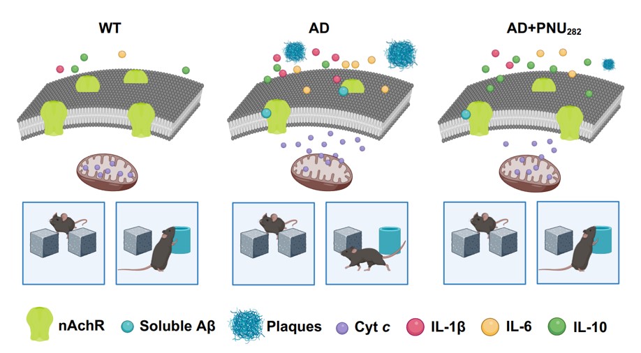 Alzheimer's disease (AD) is an age-dependent incurable neurodegenerative disorder accompanied by neuroinflammation, amyloid accumulation, and memory impairment. It begins decades before the first clinical symptoms appear, and identifying early biomarkers is key for developing disease-modifying therapies. We show now in a mouse model of AD that before any amyloid deposition the brains of 1.5-month-old mice contain increased levels of pro-inflammatory cytokines IL-1β and IL-6, decreased levels of nicotinic acetylcholine receptors (nAChRs) in the brain and brain mitochondria and increased amounts of α7 nAChR-bound Aβ1-42, along with impaired episodic memory and increased risk of apoptosis.
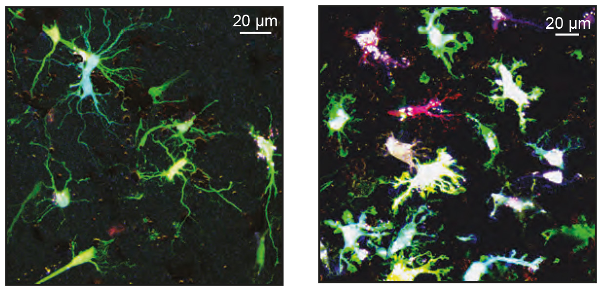 Key functions of Ca2+ signaling in rodent microglia include monitoring the brain state as well as the surrounding neuronal activity and sensing the danger or damage in their vicinity. Microglial Ca2+ dyshomeostasis is a disease hallmark in many mouse models of neurological disorders but the Ca2+ signal properties of human microglia remain unknown. We developed a novel genetically-encoded ratiometric Ca2+ indicator, targeting microglial cells in the freshly resected human tissue, organotypically cultured tissue slices and analyzed in situ ongoing Ca2+ signaling of decades-old microglia dwelling in their native microenvironment.
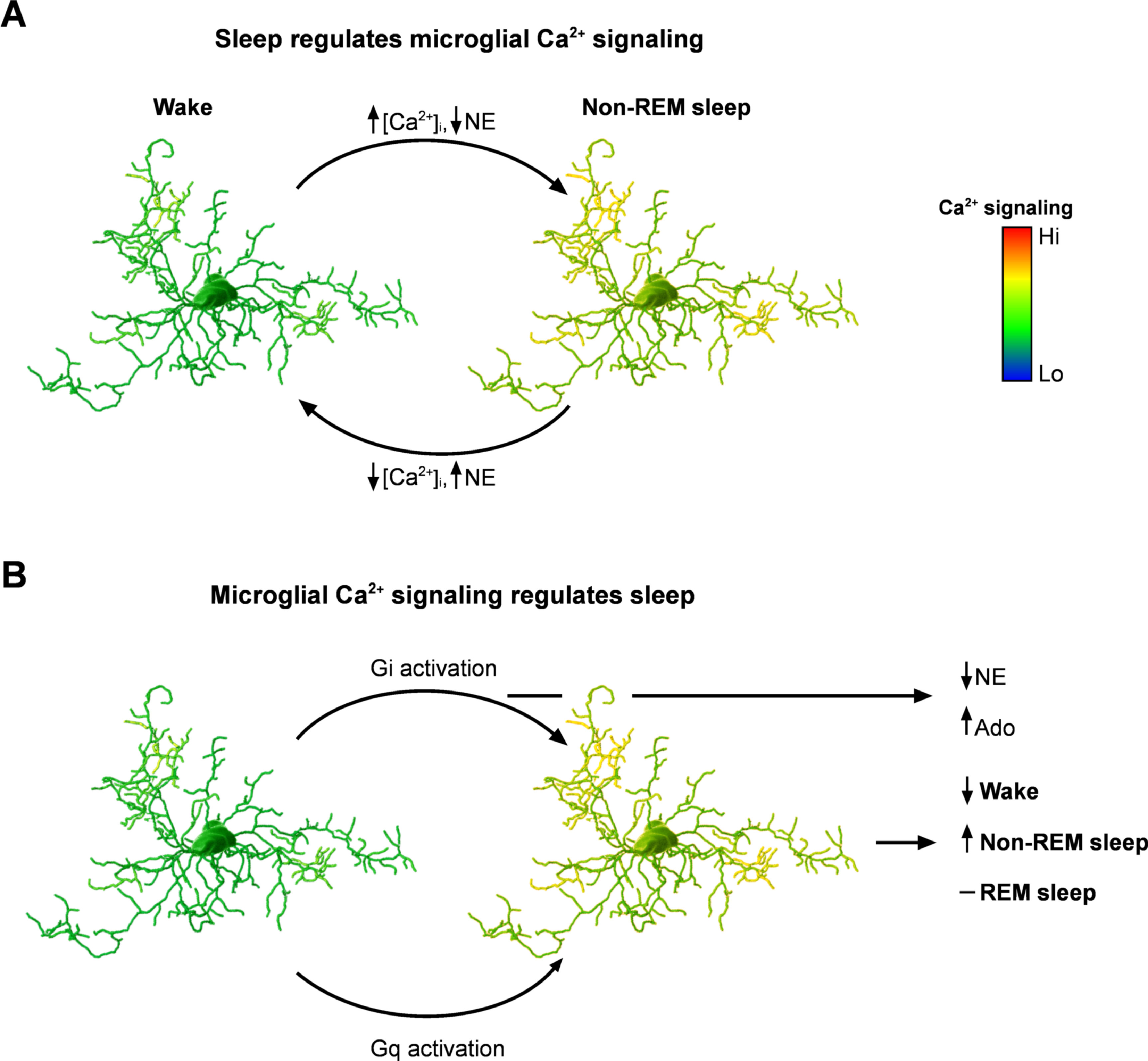 Why evolution made sleep an almost ubiquitous property of animals remains an enigma; similarly, the mechanisms regulating the sleep-wake cycle, although being extensively studied over decades remain controversial. The central role of neuroglia in sleep was proposed by Santiago Ramón y Cajal in 1895. He postulated that fine processes of astrocytes can insert in-between synaptic contacts thus limiting information transfer and instigating sleep.