Alzheimer's disease (AD) is an age-dependent incurable neurodegenerative disorder accompanied by neuroinflammation, amyloid accumulation, and memory impairment. It begins decades before the first clinical symptoms appear, and identifying early biomarkers is key for developing disease-modifying therapies. We show now in a mouse model of AD that before any amyloid deposition the brains of 1.5-month-old mice contain increased levels of pro-inflammatory cytokines IL-1β and IL-6, decreased levels of nicotinic acetylcholine receptors (nAChRs) in the brain and brain mitochondria and increased amounts of α7 nAChR-bound Aβ1-42, along with impaired episodic memory and increased risk of apoptosis.

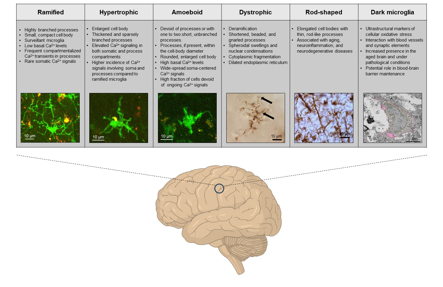
Morphotype-specific calcium signaling in human microglia Key functions of Ca2+ signaling in rodent microglia include monitoring the brain state as well as the surrounding neuronal activity and sensing the danger or damage in their vicinity. Microglial Ca2+ dyshomeostasis is a disease hallmark in many mouse models of neurological disorders but the Ca2+ signal properties of human microglia remain unknown. We developed a novel genetically-encoded ratiometric Ca2+ indicator, targeting microglial cells in the freshly resected human tissue, organotypically cultured tissue slices and analyzed in situ ongoing Ca2+ signaling of decades-old microglia dwelling in their native microenvironment.

Sleep, calcium and microglia – an (un)expected liaisonWhy evolution made sleep an almost ubiquitous property of animals remains an enigma; similarly, the mechanisms regulating the sleep-wake cycle, although being extensively studied over decades remain controversial. The central role of neuroglia in sleep was proposed by Santiago Ramón y Cajal in 1895. He postulated that fine processes of astrocytes can insert in-between synaptic contacts thus limiting information transfer and instigating sleep.

The role of intracellular calcium-store-mediated calcium signals in in vivo sensor and effector functions of microgliaUnder physiological conditions microglia, the immune sentinels of the brain, constantly monitor their microenvironment. In the case of danger, damage or cell/tissue dyshomeostasis, they react with changes in process motility, polarization, directed process movement, morphology and gene expression profile; release pro- and anti-inflammatory mediators; proliferate; and clean brain parenchyma by means of phagocytosis.

Live imaging of microglia during sleeping sickness reveals early and heterogeneous inflammatory responses

Invasion of the central nervous system (CNS) is the most serious consequence of Trypanosoma brucei infection, which causes sleeping sickness. Recent experimental data have revealed some more insights into the disease during the meningoencephalitic stage. However, detailed cellular processes befalling the CNS during the disease are poorly understood.

K+ channel-mediated retarded maturation of interneurons and its role in neurodevelopmental disordersDe novo mutations in genes encoding K+ channels are implicated in many severe neurodevelopmental disorders. Specifically, mutations in KCNA2, encoding the Shaker-type voltage-gated K+ channel Kv1.2, and KCNJ2, encoding the inwardly rectifying K+ channel Kir2.1, associate with focal and generalized epilepsies, brain atrophy, autism, ataxia and hereditary spastic paraplegia (Syrbe et al., 2015; Masnada et al., 2017; Cheng et al., 2021).

Fig for Glia Article

Microglia, the major immune cells of the brain, are functionally heterogeneous but in vivo functional properties of these cells are rarely studied at single-cell resolution. By using microRNA-9 regulated viral vectors for multicolor labeling and longitudinal in vivo monitoring of individual microglia, we followed their fate in the cortex of healthy adult mice and at the onset of amyloidosis in a mouse model of Alzheimer's disease. In wild-type mice, microglia were rather mobile (16% of the cells migrated at least once in 10–20 days) but had a low turnover as documented by low division and death rates.

Article Figure 4

Microglia arise from the yolk sac and enter the brain during early embryogenesis. Upon entry, microglia undergo in situ proliferation and eventually colonize the entire brain by the third postnatal week in mice. However, the intricacies of their developmental expansion remain unclear. Here, we characterize the proliferative dynamics of microglia during embryonic and postnatal development using complementary fate-mapping techniques. We demonstrate that the developmental colonization of the brain is facilitated by clonal expansion of highly proliferative microglial progenitors that occupy spatial niches throughout the brain.

Article Figure 6

Adult-born cells, arriving daily into the rodent olfactory bulb, either integrate into the neural circuitry or get eliminated. However, whether these two populations differ in their morphological or functional properties remains unclear. Using longitudinal in vivo two-photon imaging, we monitored dendritic morphogenesis, odor-evoked responsiveness, ongoing Ca2+ signaling, and survival/death of adult-born juxtaglomerular neurons (abJGNs). We found that the maturation of abJGNs is accompanied by a significant reduction in dendritic complexity, with surviving and subsequently eliminated cells showing similar degrees of dendritic remodeling.

Article Figure 5

The development and survival of adult-born neurons are believed to be driven by sensory signaling. Here, in vivo analyses of motility, morphology and Ca2+ signaling, as well as transcriptome analyses of adult-born juxtaglomerular cells with reduced endogenous excitability (via cell-specific overexpression of either Kv1.2 or Kir2.1 K+ channels), revealed a pronounced impairment of migration, morphogenesis, survival, and functional integration of these cells into the mouse olfactory bulb, accompanied by a reduction in cytosolic Ca2+ fluctuations, phosphorylation of CREB and pCREB-mediated gene expression.