 Using mouse olfactory bulb as a model system we compare the performance of genetically-encoded calcium sensor TN-XXL and small molecule calcium indicators; describe how to choose the right calcium indicator and how to load it into the cells of interest; discuss the use of cell type-specific markers and, finally, illustrate the application of this technique for high resolution in vivo imaging of sensory-driven neuronal activity.
For more details see:
In vivo functional imaging of the olfactory bulb at single cell resolution, S, Fink, Y Kovalchuk, R Homma, B Schwendele, S Direnberger, LB Cohen, O Griesbeck, and O Garaschuk, Neuronal Network Analysis (in Press)
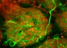
 Juxtaglomerular neurons represent one of the largest cellular populations in the mammalian olfactory bulb yet their role for signal processing remains unclear. We used two-photon imaging and electrophysiological recordings to clarify the in vivo properties of these cells and their functional organization in the juxtaglomerular space. Juxtaglomerular neurons coded for many perceptual characteristics of the olfactory stimulus such as (1) identity of the odorant, (2) odorant concentration, (3) odorant onset, and (4) offset.
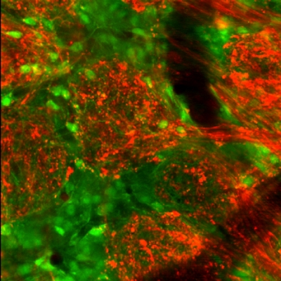
 We describe a technique utilizing Tomato lectin conjugated with green or red fluorescent dye for fast and reliable in vivo labeling of microglia. This protocol enables high resolution imaging of microglial cells in wild type/mutant mice of any age, and in mouse models of Alzheimer’s disease. The labeling does not disturb the functional properties of microglia or the surrounding neurons and is preserved in fixed tissue used for post-hoc immunostaining.
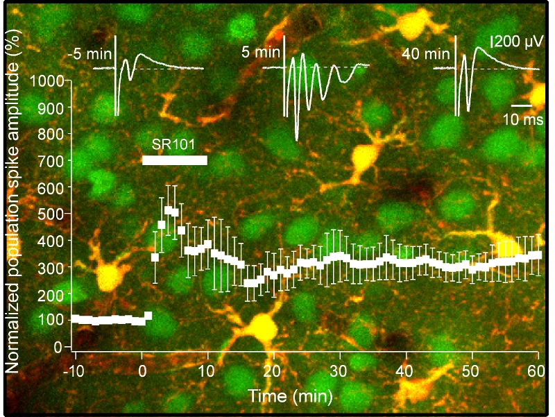 SR101 turned out to be a highly bioactive molecule. When tested in acutely prepared hippocampal slices this substance markedly increased the excitability of neuronal tissue (Fig. 1B-F, (Fink et al., 2011)). Bath application of SR101 induced a 5-6 fold increase in the amplitude of a synaptically-evoked population spike caused by the stimulation of Schaffer collaterals and provoked a synchronized repetitive firing of neurons at a frequency of 130-200 Hz. The SR101-evoked hyperexcitability developed rapidly (the maximal effect was reached during the first 5 min of drug application) and persisted for at least 40-60 min after the wash-out of the drug. Field EPSPs recorded in the stratum radiatum of the CA1 region also underwent long lasting potentiation.
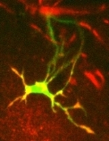
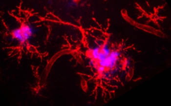
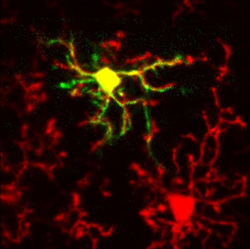 In the healthy adult brain microglia, the main immune-competent cells of the CNS, have a distinct (so-called “resting”, see image) phenotype. Resting microglia can only be studied in vivo since any isolation of brain tissue inevitably triggers microglial activation. Using in vivo two-photon imaging we obtained a first direct insight into Ca2+ signaling in resting cortical microglia.