In vivo characterization of functional states of cortical microglia during peripheral inflammation.
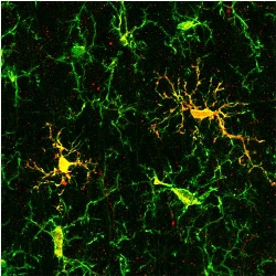 Peripheral inflammation is known to trigger a mirror inflammatory response in the brain, involving brain's innate immune cells - microglia. However, the functional phenotypes, which these cells adopt in the course of peripheral inflammation, remain obscure. In vivo two-photon imaging of microglial Ca2+ signaling as well as process motility reveals two distinct functional states of cortical microglia during a lipopolysaccharide-induced peripheral inflammation: an early "sensor state" characterized by dramatically increased intracellular Ca signaling but ramified morphology and a later "effector state" characterized by slow normalization of intracellular Ca2+ signaling but hypertrophic morphology, substantial IL-1β production in a subset of cells as well as increased velocity of directed process extension and loss of coordination between individual processes. Thus, lipopolysaccharide-induced microglial Ca2+ signaling might represent the central element connecting receptive and executive functions of microglia.
Recommended in Faculty Opinions:
Shih A, Tieu T and Coelho-Santos V: Faculty Opinions Recommendation of [Riester K et al., Brain Behav Immun 2020 87:243-255]. In Faculty Opinions, 09 Jun 2021; 10.3410/f.737086570.793585866
see more: https://www.sciencedirect.com/science/article/pii/S0889159119312942?via%3Dihub
- 113 views