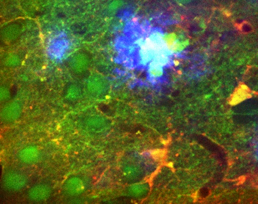
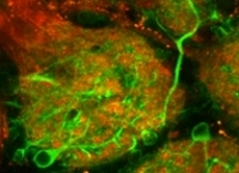
 Using mouse olfactory bulb as a model system we compare the performance of genetically-encoded calcium sensor TN-XXL and small molecule calcium indicators; describe how to choose the right calcium indicator and how to load it into the cells of interest; discuss the use of cell type-specific markers and, finally, illustrate the application of this technique for high resolution in vivo imaging of sensory-driven neuronal activity.
For more details see:
In vivo functional imaging of the olfactory bulb at single cell resolution, S, Fink, Y Kovalchuk, R Homma, B Schwendele, S Direnberger, LB Cohen, O Griesbeck, and O Garaschuk, Neuronal Network Analysis (in Press)

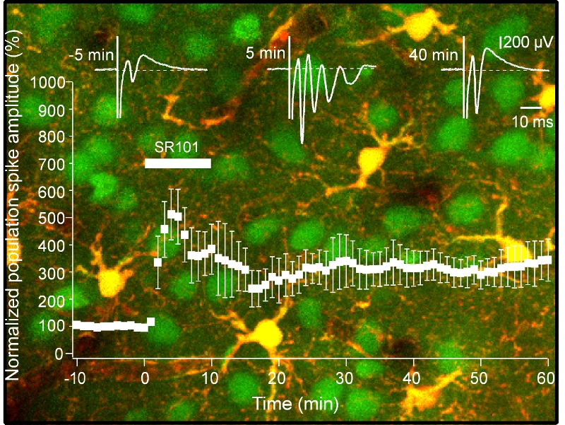 SR101 turned out to be a highly bioactive molecule. When tested in acutely prepared hippocampal slices this substance markedly increased the excitability of neuronal tissue (Fig. 1B-F, (Fink et al., 2011)). Bath application of SR101 induced a 5-6 fold increase in the amplitude of a synaptically-evoked population spike caused by the stimulation of Schaffer collaterals and provoked a synchronized repetitive firing of neurons at a frequency of 130-200 Hz. The SR101-evoked hyperexcitability developed rapidly (the maximal effect was reached during the first 5 min of drug application) and persisted for at least 40-60 min after the wash-out of the drug. Field EPSPs recorded in the stratum radiatum of the CA1 region also underwent long lasting potentiation.
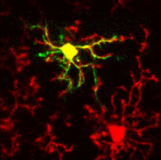 In the healthy adult brain microglia, the main immune-competent cells of the CNS, have a distinct (so-called “resting”, see image) phenotype. Resting microglia can only be studied in vivo since any isolation of brain tissue inevitably triggers microglial activation. Using in vivo two-photon imaging we obtained a first direct insight into Ca2+ signaling in resting cortical microglia.