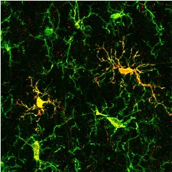
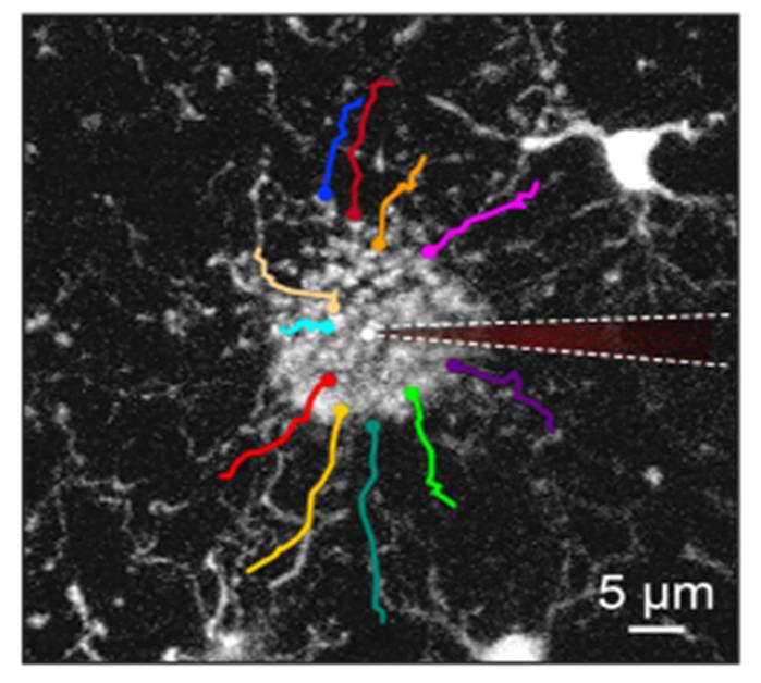
 Throughout the lifespan, microglia, the primary innate immune cells of the brain, fulfill a plethora of homeostatic as well as active immune defense functions, and their aging-induced dysfunctionality is now considered as a key trigger of aging-related brain disorders. Recent evidence suggests that both organism’s sex and age critically impact the functional state of microglia but in vivo determinants of such state(s) remain unclear.
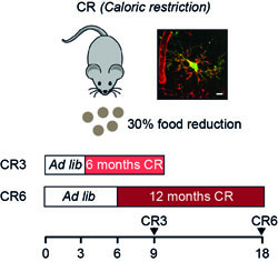
Therefore, we analyzed in vivo the sex-specific functional states of microglia in young adult, middle aged and old wild type mice by means of multicolor two-photon imaging, using the microglial Ca2 + signaling and directed process motility as main readouts.
read more: https://www.frontiersin.org/articles/10.3389/fimmu.2020.00750/full
 Neuronal hyperactivity is the emerging functional hallmark of Alzheimer’s disease (AD) in both humans and different mouse models, mediating an impairment of memory and cognition. The mechanisms underlying neuronal hyperactivity remain, however, elusive. In vivo Ca2+ imaging of somatic, dendritic, and axonal activity patterns of cortical neurons revealed that both healthy aging and AD-related mutations augment neuronal hyperactivity. The AD-related enhancement occurred even without amyloid deposition and neuroinflammation, mainly due to presenilin-mediated dysfunction of intracellular Ca2+ stores in presynaptic boutons, likely causing more frequent activation of synaptic NMDA receptors.